Transmission of crAsspahge in the microbiome

Update! This work has been published in Nature Communications.
Siranosian, B.A., Tamburini, F.B., Sherlock, G. et al. Acquisition, transmission and strain diversity of human gut-colonizing crAss-like phages. Nat Commun 11, 280 (2020). https://doi.org/10.1038/s41467-019-14103-3

Big questions in the microbiome field surround the topic of microbiome acquisition. Where do we get our first microbes from? What determines the microbes that colonize our guts form birth, and how do they change over time? What short and long term impacts do these microbes have on the immune system, allergies or diseases? What impact do delivery mode and breastfeeding have on the infant microbiome?

A key finding from the work was that mothers and infants often share identical or nearly identical crAssphage sequences, suggesting direct vertical transmission. Also, I love heatmaps.

As you might expect, a major source for microbes colonizing the infant gut is immediate family members, and the mother is thought to be the major source. Thanks to foundational studies by Bäckhed, Feretti, Yassour and others (references below), we now know that infants often acquire the primary bacterial strain from the mother’s microbiome. These microbes can have beneficial capabilities for the infant, such as the ability to digest human milk oligosaccharides, a key source of nutrients in breast milk.

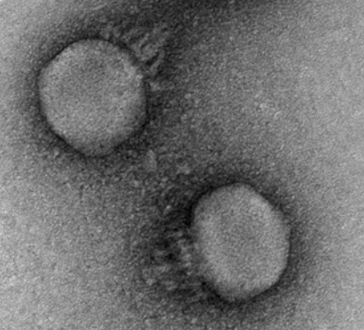
The microbiome isn’t just bacteria – phages (along with fungi and archaea to a smaller extent) play key roles. Phages are viruses that predate on bacteria, depleting certain populations and exchanging genes among the bacteria they infect. Interestingly, phages were previously shown to display different inheritance patterns than bacteria, remaining individual-specific between family members and even twins (Reyes et al. 2010). CrAss-like phages are the most prevalent and abundant group of phages colonizing the human gut, and our lab was interested in the inheritance patterns of these phages.

We examined publicly available shotgun gut metagenomic datasets from two studies (Yassour et al. 2018, Bäckhed et al. 2015), containing 134 mother-infant pairs sampled extensively through the first year of life. In contrast to what has been observed for other members of the gut virome, we observed many putative transmission events of a crAss-like phage from mother to infant. The key takeaways from our research are summarized below. You can also refer my poster from the Cold Spring Harbor Microbiome meeting for the figures supporting these points. We hope to have a new preprint (and hopefully a publication) on this research out soon!

  1. CrAssphage is not detected in infant microbiomes at birth, increases in prevalence with age, but doesn’t reach the level of adults by 12 months of age.
  2. Mothers and infants share nearly identical crAssphage genomes in 40% of cases, suggesting vertical transmission.
  3. Infants have reduced crAssphage strain diversity and typically acquire the mother’s dominant strain upon transmission.
  4. Strain diversity is mostly the result of neutral genetic variation, but infants have more nonsynonymous multiallelic sites than mothers.
  5. Strain diversity varies across the genome, and tail fiber genes are enriched for strain diversity with nonsynonymous variants.
  6. These findings extend to crAss-like phages. Vaginally born infants are more likely to have crAss-lke phages than those born via C-section.

References
1. Reyes, A. et al. Viruses in the faecal microbiota of monozygotic twins and their mothers. Nature 466, 334–338 (2010).
2. Yassour, M. et al. Strain-Level Analysis of Mother-to-Child Bacterial Transmission during the First Few Months of Life. Cell Host & Microbe 24, 146-154.e4 (2018).
3. Bäckhed, F. et al. Dynamics and Stabilization of the Human Gut Microbiome during the First Year of Life. Cell Host & Microbe 17, 690–703 (2015).
4. Ferretti, P. et al. Mother-to-Infant Microbial Transmission from Different Body Sites Shapes the Developing Infant Gut Microbiome. Cell Host & Microbe 24, 133-145.e5 (2018).

What is crAssphage?

CrAssphage is a like mystery novel full of surprises. First described in 2014 by Dutilh et al., crAssphage acquired it’s (rather unfortunate, given that it colonizes the human intestine) name from the “Cross-Assembly” bioinformatics method used to characterize it. CrAssphage interests me because it’s prevalent in up to 70% of human gut microbiomes, and can make up the majority of viral sequencing reads in a metagenomics experiment. This makes it the most successful single entity colonizing human microbiomes. However, no health impacts have been demonstrated from having crAssphage in your gut – several studies (Edwards et al. 2019) have turned up negative.

Electron micrograph of a representative crAssphage, from Shkoporov et al. (2018). This phage is a member of the Podoviridae family and infects Bacteroides Intestinalis.

CrAssphage was always suspected to predate on species of the Bacteroides genus based on evidence from abundance correlation and CRISPR spacers. However, the phage proved difficult to isolate and culture. It wasn’t until recently that a crAssphage was confirmed to infect Bacteroides intestinalis (Shakoporov et al. 2018). They also got a great TEM image of the phage! With crAssphage successfully cultured in the lab, scientists have begun to answer fundamental questions about its biology. The phage appears to have a narrow host range, infecting a single B. intestinalis strain and not others or other species. The life cycle of the phage was puzzling:

“We can conclude that the virus probably causes a successful lytic infection with a size of progeny per capita higher than 2.5 in a subset of infected cells (giving rise to a false overall burst size of ~2.5), and also enters an alternative interaction (pseudolysogeny, dormant, carrier state, etc.) with some or all of the remaining cells. Overall, this allows both bacteriophage and host to co-exist in a stable interaction over prolonged passages. The nature of this interaction warrants further investigation.” (Shakoporov et al. 2018)

Further investigation showed that crAssphage is one member of an extensive family of “crAss-like” phages colonizing the human gut. Guerin et al. (2018) proposed a classification system for these phages, which contains 4 subfamilies (Alpha, Beta, Delta and Gamma) and 10 clusters. The first described “prototypical crAssphage” belongs to the Alpha subfamily, cluster 1. It struck me how diverse these phages are – different families are less than 20% identical at the protein level! When all crAss-like phages are considered, it’s estimated that up to 100% of individuals cary at least one crAss-like phage, and most people cary more than one.

Given the high prevalence of crAss-like phages and their specificity for the human gut, they do have an interesting use as a tracking device for human sewage. DNA from crAss-like phages can be used to track waste contamination into water, for example (Stachler et al. 2018). In a similar vein, our lab has used crAss-like phages to better understand how microbes are transmitted from mothers to newborn infants. The small genome sizes (around 100kb) and high prevalence/abundance make these phages good tools for doing strain-resolved metagenomics. Trust me, you’d much rather do genomic assembly and variant calling on a 100kb phage genome than a 3Mb bacterial genome!

Research into crAss-like phages is just beginning, and I’m excited to see what’s uncovered in the future. What are the hosts of the various phage clusters? How do these phages influence gut bacterial communities? Do crAss-like phages exclude other closely related phages from colonizing their niches, leading to the low strain diversity we observe? Can crAss-like phgaes be used to engineer bacteria in the microbiome, delivering precise genetic payloads? This final question in the most interesting to me, given that crAss-like phages seem relatively benign to humans, yet incredibly capable of infecting our microbes.

References
1.Dutilh, B. E. et al. A highly abundant bacteriophage discovered in the unknown sequences of human faecal metagenomes. Nature Communications 5, 4498 (2014).
2.Edwards, R. A. et al. Global phylogeography and ancient evolution of the widespread human gut virus crAssphage. Nature Microbiology 1 (2019). doi:10.1038/s41564-019-0494-6
3.Guerin, E. et al. Biology and Taxonomy of crAss-like Bacteriophages, the Most Abundant Virus in the Human Gut. Cell Host & Microbe 0, (2018).
4.Shkoporov, A. N. et al. ΦCrAss001 represents the most abundant bacteriophage family in the human gut and infects Bacteroides intestinalis. Nature Communications 9, 4781 (2018).
5.Stachler, E., Akyon, B., de Carvalho, N. A., Ference, C. & Bibby, K. Correlation of crAssphage qPCR Markers with Culturable and Molecular Indicators of Human Fecal Pollution in an Impacted Urban Watershed. Environ. Sci. Technol. 52, 7505–7512 (2018).

Metagenome Assembled Genomes enhance short read classification

In the microbiome field we struggle with the fact that reference databases are (sometimes woefully) incomplete. Many gut microbes are difficult to isolate and culture in the lab or simply haven’t been sampled frequently enough for us to study. The problem is especially bad when studying microbiome samples from non-Western individuals.

To subvert the difficulty in culturing new organisms, researchers try to create new reference genomes directly from metagenomic samples. This typically uses metagenomic assembly and binning. Although you most likely end up with a sequence that isn’t entirely representative of the organism, these Metagenome Assembled Genomes (MAGs) are a good place to start. They provide new reference genomes for classification and association testing, and start to explain what’s in the microbial “dark matter” from a metagenomic sample.

2019 has been a good year for MAGs. Three high profile papers highlighting MAG collections were published in the last few months[1,2,3]. The main idea in each of them was similar – gather a ton of microbiome data, assemble and bin contigs, filter for quality and undiscovered genomes, do some analysis of the results. My main complaint about all three papers is that they use reduced quality metrics, not following the standards set in Bowers et al. (2017). They rarely find 16S rRNA sequences in genomes called “high quality,” for example.

Comparing the datasets, methods, and results from the three MAG studies. This table was compiled by Yiran Liu during her Bhatt lab rotation.

After reading the three MAG papers, Nayfach et al. (2019) is my favortie. His paper does the most analysis into what these new genomes _mean_, including a great finding presented in Figure 4. These new references assembled from metagenomes can help explain why previous studies looking for associations between the microbiome and disease have come up negative. This can also help explain why microbiome studies have been difficult to replicate. If a significant association is hiding in these previously unclassified genomes, a false positive association could easily look significant because everything is tested with relative abundance.

In the Bhatt lab, we were interested in using these new MAG databases to improve classification rates in some samples from South African individuals. First we had to build a Kraken2 database for the MAG collections. If you’re interested in how to do this, I have an instructional example over at the Kraken2 classification GitHub. For samples from Western individuals, the classification percentages don’t increase much with MAG databases, in line with what we would expect. For samples from South African individuals, the gain is sizeable. We see the greatest increase in classification percentages by using the Almeida et al. (2019) genomes. This collection is the largest, and may represent a sensitivity/specificity tradeoff. The percentages represented below for MAG databases are calculated as the total classifies percentages when the unclassified reads from our standard Kraken2 database are run through the MAG database.

Classification percentages on samples from Western individuals. We’re already doing pretty good without the MAG database.

Classification percentages on non-Western individuals. MAGs add a good amount here. Data collected and processed by Fiona Tamburini.


References
1.Nayfach, S., Shi, Z. J., Seshadri, R., Pollard, K. S. & Kyrpides, N. C. New insights from uncultivated genomes of the global human gut microbiome. Nature 568, 505 (2019).
2.Pasolli, E. et al. Extensive Unexplored Human Microbiome Diversity Revealed by Over 150,000 Genomes from Metagenomes Spanning Age, Geography, and Lifestyle. Cell 0, (2019).
3.Almeida, A. et al. A new genomic blueprint of the human gut microbiota. Nature 1 (2019). doi:10.1038/s41586-019-0965-1
4.Bowers, R. M. et al. Minimum information about a single amplified genome (MISAG) and a metagenome-assembled genome (MIMAG) of bacteria and archaea. Nature Biotechnology 35, 725–731 (2017).

Race report: China Peak Enduro 2019

Rocky, rocky China Peak

China Peak was a rocky, rocky race. Photo creadit Scott McClain.

I raced the China Peak Enduro mountain bike race last weekend with Nick, Catherine, Peter Bai and Kate (visiting MTB rider from MIT who casually placed 3rd in PRO women). We all survived and even managed to have a good time! I placed toward the bottom of the pack in the sport category, but I’m going to blame it on a flat forcing me to run with my bike though the second half of stage 3.

If you’ve never seen an enduro race and want some context for the stage 3 rock garden I mention, check out this video. We’re talking mandatory hucks off rock drops and sandy blown-out corners wherever you look. It’s both a mental and physical game – the course gets easier the more momentum you carry through the features, but that is so much easier said than done. The China Peak Enduro consisted of 5 timed downhill segments, with pedal or ski lift transfers between them. I raced in the sport category, so I only did stages 1-4. Friday was a practice day where we sessioned the hard segments and tried to pick out the right lines. The race then began early on Saturday morning.

Stage 1: We had a long (45-60min) but easy pedal to the top of China Peak for the start of stage 1. This downhill section was flowey and fun. You had to trust the blown-out, sandy berms that they would hold you as you whipped around them, and be ready to put a foot out when they didn’t. There was one part of this stage that was an absolute mud pit. You had to stay on the slippery bridge or risk putting your bike into a foot of mud!
Time: 0:05:18 Position after this stage: 16/23

Easy pedal up to the top of stage 2.

Stage 2: Another easier, flow stage with a few rocky sections. The most challenging part was the bottom few corners that were basically sand pits with large rocks in them. How people took these at speed, I have no idea. There was no support for your tires. Your best bet was to dive in from the high line and try to slide your back wheel around.
Time: 0:06:00 Position after this stage: 14/23

Chair lift ride to the top of stage 3.

Stage 3: This stage was broken up into 4 sections: A rocky and fast section first, a pedal heavy middle, the rock garden (perhaps the hardest single feature of the whole day), and a pedal heavy end. Nick and I spent a while in practice trying to pick out the best lines through the rock garden, but I still hadn’t ridden it clean. I was nervous about this section, but hoped the race day adrenaline would carry me though. By this time I was warmed up and riding well – the top of stage 3 flew by and I was charging off the rock drops.
A few hundred feet before the dreaded rock garden, I hit something sharp and cut a slash in my front tire. In a few seconds my pressure and sealant was gone – I knew this wasn’t a trailside repair. I made the decision to run the rest of the course as fast as possible. The other option was putting in a tube, which would have cost more time in the end I think. So up on the shoulder the 30lb MTB goes, and I start hopping down the rock garden (cue massive heckling from the onlookers). I was passed by a few riders from behind before the finish. Secretly relieved I didn’t break myself on the rock garden? Maybe.

Time: 0:15:32 (the single worst stage 3 time) Position: 22/23

Chair lift ride to the top of stage 4.

Stage 4: Long, steep rock slabs was the theme of this stage. Try and maintain good bike position while dropping off of boulders… I was surprised how quickly my upper body tired out. You might not consider a 9 minute stage an endurance event, but I was feeling beat. I put down a respectable time on this stage, even managing to pass the guy in front of me. Sadly, it wasn’t enough to make up for the several minutes I lost on stage 3. On the flat sprint to the finish, I managed to snap my chain! My bike is in need of some serious love after the weekend.
Time: 0:08:58 Final position: 21/23

Overall, I had a great time racing and camping with the Stanford crew, and got to work on my skills a lot at China Peak. This is my first proper downhill event of the season, so I know I have a lot to work on. We’re racing the Mt. Shasta Enduro in a week and a half, so that will be the next test!